Ask why muscle fades after 60 and you will hear about protein intake, anabolic resistance, and lost training stimulus. All of that is real. But it does not explain why aged muscle often feels underpowered before it looks visibly smaller — why the first loss is speed, grip, and the ability to catch yourself on a stumble.
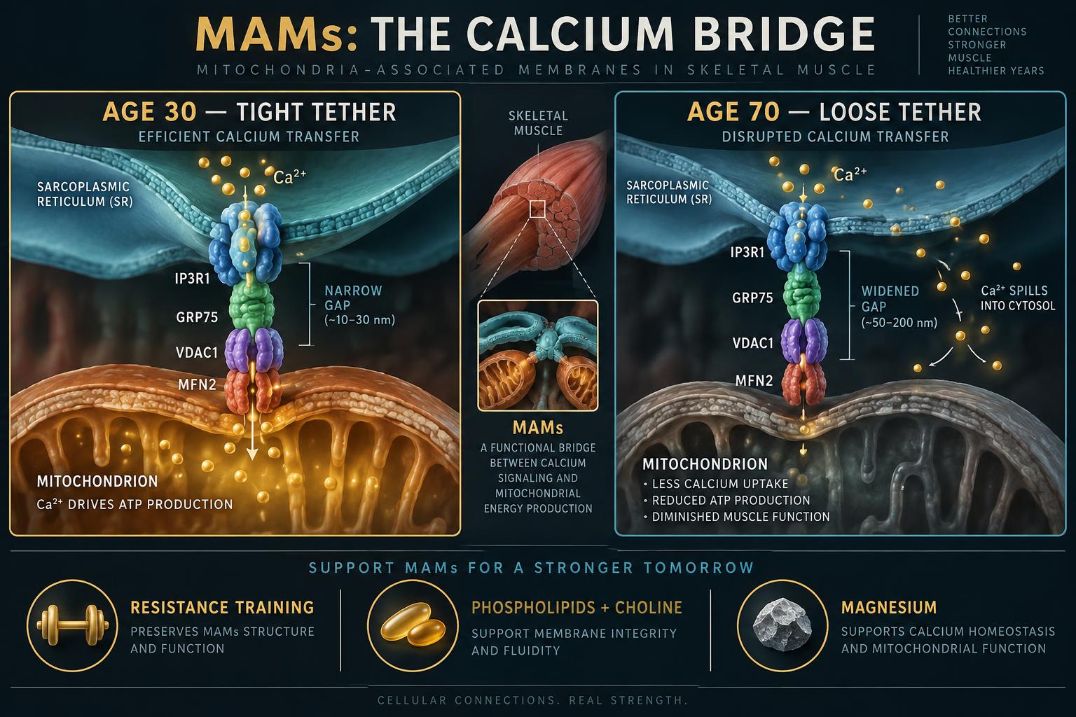
A structural answer is emerging from electron-microscopy and ultrastructural work on aged skeletal muscle. The problem is not only how much tissue is left. It is that the physical contact points where the sarcoplasmic reticulum hands calcium directly to mitochondria — the mitochondria-associated membranes, or MAMs — pull apart with age. When that gap widens, the contraction signal and the energy response fall out of sync.
Contents(13 sections)
Key takeaways
MAMs are narrow tethered junctions (roughly 10–30 nm) where the sarcoplasmic reticulum releases calcium straight into mitochondria to switch on ATP production.
Calcium is not just the contraction trigger; inside the mitochondrion it is the accelerator pedal for the enzymes that make energy.
Aged muscle shows widened, fewer, and less organized SR–mitochondrial contacts, with reduced tethering proteins — a structural lesion independent of total protein intake.
The consequence is mistimed energy: calcium leaks into the cytosol instead of reaching the mitochondrial matrix, so power output falls before visible mass does.
Resistance training is the strongest known stimulus for restoring contact-site architecture; membrane phospholipid supply and magnesium sufficiency support it.
The Primer
What a MAM actually is
Two structures sit side by side in every muscle fiber. The sarcoplasmic reticulum is the storage tank that dumps calcium to trigger contraction. The mitochondrion is the power plant that pays for it. They are not floating independently — they are physically tied together at specialized junctions only a few tens of nanometers wide.
Those junctions are the MAMs. Their job is handoff. When the SR releases calcium, a portion of it is delivered at close range, straight into the mitochondrion, rather than diffusing through the cell and getting diluted.
Why the handoff matters
Calcium inside a mitochondrion is a command, not a waste product. It activates the dehydrogenase enzymes that feed the electron transport chain, which raises ATP output within milliseconds of a contraction starting.
That is the elegance of the design: the same ion that tells the muscle to contract also tells the mitochondria to pay for the contraction. Demand and supply are signaled by one molecule, at one moment, because the two membranes are touching.
What goes wrong with age
In aged muscle, the gap between the two membranes widens, the number of well-formed contacts drops, and the proteins that hold the tether in place become less abundant. The tank still releases calcium, and the power plant still works — but the message no longer arrives cleanly.
Calcium that should have entered the mitochondrion lingers in the cytosol instead. That has two costs: mitochondria are under-stimulated exactly when they are needed most, and the cytosol carries a chronic calcium load that drives proteolysis and low-grade stress signaling in the fiber.
What this changes about the plan
It does not overturn the fundamentals. Protein and resistance training remain the base of the work. What it changes is the explanation for a common frustration: older adults who train and eat well can still feel underpowered, because rebuilding contractile protein does not automatically rebuild the architecture that times the energy delivery.
Structure responds to load, slowly. That is an argument for consistency and for training that demands speed and force, not only volume.
The Deep Dive
The tether complex
The canonical bridge is a three-protein assembly: an IP3 receptor (IP3R1) on the reticulum membrane, the outer-mitochondrial channel VDAC1, and the cytosolic chaperone GRP75 linking the two. Mitofusin-2 (MFN2), best known for mitochondrial fusion, also functions here as a structural tether that sets and maintains the inter-membrane distance.
In skeletal muscle the same principle operates at the triad and at SR–mitochondrial contacts adjacent to it, where the ryanodine receptor dominates release. The mitochondrial calcium uniporter (MCU) is the entry gate on the inner membrane, and it has low affinity for calcium — which is precisely why proximity is non-negotiable. MCU only sees the high-concentration microdomain created within nanometers of a release site. Widen the gap and the uniporter is effectively blind to a release that still happens normally.
Tier II · Members only · Free
Continue into the deep dive
Tier I is free and always open. Tier II — the full mechanisms, dosing detail, nuance, and citations — unlocks with a free Vital Codex membership.
- Every Tier II deep dive across all hubs
- Saved reading progress across articles
- Member knowledge modules and their quizzes
- The Dispatch, if you opt in
Frequently asked
What does MAM stand for?
Mitochondria-associated membranes — the narrow, tethered contact sites where the endoplasmic or sarcoplasmic reticulum physically touches mitochondria and transfers calcium and lipids directly between them.
Is this the real cause of sarcopenia?
It is one contributing mechanism, and a structural one that the protein-and-training model does not fully explain. Sarcopenia remains multifactorial: training stimulus, protein sufficiency, hormonal signaling, inflammation, and mitochondrial capacity all contribute. The MAM literature adds a plausible explanation for why power declines faster than mass.
Can MAM function be measured in a person?
Not routinely. Contact-site geometry requires electron microscopy on a biopsy. Practical functional proxies are grip strength, chair-rise speed, and peak power output, all of which decline earlier than lean mass on a scan.
Does any supplement fix the calcium bridge?
No supplement has been shown to restore contact-site architecture. Mechanical loading is the intervention with a plausible structural effect. Phospholipid precursors, omega-3s, magnesium, and creatine support the surrounding system and the energy buffer, which is a genuinely different claim.
Why do eggs keep appearing in muscle-aging protocols?
Because whole eggs supply leucine-rich protein and phosphatidylcholine in the same food, and membrane repair needs both. Free-range and pasture-raised eggs are worth the premium here: the fat fraction of the yolk is the part doing the membrane work, and pasture access measurably improves its fatty-acid profile and antioxidant content.
How long before training changes anything structural?
Contractile and metabolic adaptations appear within four to six weeks. Ultrastructural reorganization is slower and is documented over months of consistent loading. This is a multi-year maintenance project, not a protocol with an endpoint.
Research Notes & Sources(expand)
Senapati U, Kar BP, Pani S, et al. "Aging induced structural alterations in SR-Mitochondria interaction in skeletal muscle: Emerging insights." arXiv preprint arXiv:2606.17457 (2026). Review of ultrastructural evidence that SR–mitochondrial contact geometry and tethering-protein expression degrade with age in skeletal muscle.
Holt KB, Zurita C, Teryoshin L, et al. "Diffusive Spreading Across Dynamic Mitochondrial Network Architectures." arXiv preprint arXiv:2506.05643 (2025). Temporal-network modeling showing that fusion–fission dynamics, not organelle count, govern delivery of metabolites and signals within a cell.
Fahimi P, Lynch M, Matta CF. "Decoding the Hot-Mitochondrion Paradox." arXiv preprint arXiv:2507.16824 (2025). Non-equilibrium thermodynamic treatment of localized energy dissipation at the inner mitochondrial membrane.
Editorial note: this article is educational synthesis of published research, including preprints that have not completed peer review. It is not medical advice and does not establish a clinician–patient relationship. Supplement and training decisions in the presence of cardiac, renal, or neuromuscular disease require a qualified clinician.
Continue exploring: Muscle & Aging — Hypertrophy, sarcopenia prevention, and structural health.
Explore Muscle & Aging